Wednesday, April 22, 2009
PITCH ARTIFACTS IN MULTISECTION SCANNING
As helical pitch increases, the number of detector rows intersecting the image plane per rotation increases and the number of "vanes" in the windmill artifact increases. One of the benefits of z-filter interpolators is that they reduce the severity of windmill artifacts, especially when the image reconstruction width is wider than the detector acquisition width. Artifacts may also be slightly reduced by using noninteger pitch values relative to detector acquisition width, such as pitches of 3.5 or 4.5 on a four-section scanner. This is because z-axis sampling density is optimized for noninteger pitches.
PITCH EFFECT
Monday, April 20, 2009
BOLUS TRACKING
The newer MDCT scanners have shorter scan times it was necessary to re-evaluate the injection protocols for CTA. Bolus tracking is a method that individualizes the timing of contrast media (CM) delivery to a region of interest (ROI). An injection of CM is tracked or observed until it reaches the desired Hounsfield Units (HU) threshold for the ROI. When that threshold is met wait a few seconds and then scan to obtain the optimal image enhancement.
This method of imaging is used primarily to produce images of arteries, such as the aorta, pulmonary artery, cerebral and carotid arteries. The image shown below illustrates this technique on a sagittal MPR (multiplanar reformat). The image is demonstrating the blood flow through an abdominal aortic aneurysm or AAA. The bright white on the image is the contrast. You can see the lumen of the aorta in which the contrast is contained, surrounded by a grey "sack", which is the aneurysm. Images acquired from a bolus track, can be manipulated into a MIP (maximum intensity projection) or a volume rendered image.
This method of imaging is used primarily to produce images of arteries, such as the aorta, pulmonary artery, cerebral and carotid arteries. The image shown below illustrates this technique on a sagittal MPR (multiplanar reformat). The image is demonstrating the blood flow through an abdominal aortic aneurysm or AAA. The bright white on the image is the contrast. You can see the lumen of the aorta in which the contrast is contained, surrounded by a grey "sack", which is the aneurysm. Images acquired from a bolus track, can be manipulated into a MIP (maximum intensity projection) or a volume rendered image.
MAXIMUM INTENSITY PROJECTION
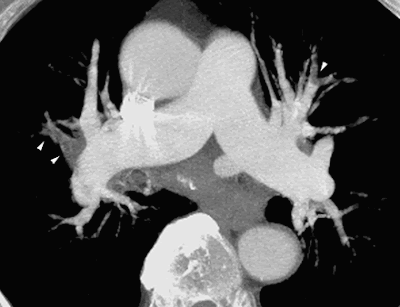
Maximum Intensity Projection (MIP) is a post processing technique that reconstructs an image by selecting the highest value pixels along any arbitrary line through the data set and exhibiting only those pixels. It creates a 3-D image from multislice 2-D data sets; it's the simplest form of 3-D imaging. MIP images are widely used in CTA because they can be reconstructed very quickly.
SEGMENTATION
Segmentation is a step in processing an object model into a simulated 3-D image. Segmentation is a processing technique used to identify the structure of interest in a given scene. It determines which voxels are a part of the object and should be displayed and which are not and should be discarded.
SEGMENTATION
ARTIFACTS IN SPIRAL CT
The following artifacts are listed on this blog.
Misregistration
Scalloping
Banding
Stair-Stepping
Pitch Effect
Misregistration
Scalloping
Banding
Stair-Stepping
Pitch Effect
MISREGISTRATION ARTIFACTS
Patient motion can cause misregistration artifacts, which usually appear as shading or streaking in the reconstructed image. Steps can be taken to prevent voluntary motion, but some involuntary motion may be unavoidable during body scanning. However, there are special features on some scanners designed to minimize the resulting artifacts.
MISREGISTRATION ARTIFACTS
 Motion artifact simulating the appearance of dissection. Postcontrast image through the ascending aorta reveals an irregular line extending across the aorta (curved arrow), simulating aortic dissection. This line was not seen on images immediately above and below this level (not shown). Motion artifacts are most prevalent and severe near the base of the heart. They may be recognized by their non-anatomic configuration and their inconstant appearance on sequential images.
SCALLOPING ARTIFACTS
Scalloping Artifact is due to the fact that the slice sensitivity profile (SSP) is increased in spiral CT so that partial volume artifacts also become stronger. Scalloping can occur in skull CTs, particularly in slice positions in which the skull diameter quickly changes its axial direction. This image error can be corrected by reducing the pitch factors.
Unable to locate any Computed Tomography (CT) Scalloping Artifact Images .
Unable to locate any Computed Tomography (CT) Scalloping Artifact Images .
SCALLOPING ARTIFACTS
BANDING ARTIFACTS
Band artifacts are produced when the projection reading of a single channel or a group of channels consistently deviate from the truth. That can be the result of defective detector cells of DAS (Data Acquisition System) channels, deficiencies in system calibration, or a suboptimal image-generation process. This is predominately a third-generation CT scanner phenomenon. The detector channel reading is always mapped to a straight line that is at a fixed distance to the isocenter of the system, a defective reading forms a ring pattern during the back-projection process.
Note: Band or Ring Artifacts are produced in the same way.
Note: Band or Ring Artifacts are produced in the same way.
Sunday, April 19, 2009
STAIR-STEP ARTIFACT
STAIR STEP ARTIFACTS
STAIR-STEP ARTIFACT
Stair step artifacts appear around the edges of structures in multiplanar and three-dimensional reformatted images when wide collimations and nonoverlapping reconstruction intervals are used. they are less severe with helical scanning, which permits reconstruction of overlapping sections without the extra dose to the patient that would occur if overlapping axial scans were obtained. Stair step artifacts are virtually eliminated in multiplanar and three-dimensional reformatted images from thin-section data obtained with today's multisection scanners.
SPIRAL CT PITCH EFFECT
PITCH EFFECT:
CT pitch is generally defined as the table travel per rotation divided by the collimation of the x-ray beam. This beam-pitch definition can also be refered to as table travel per rotation divided by effective detector row thickness. Thus, a beam-pitch of 1.0 facilitates an acquisition with no overlap or gap, a beam-pitch of less than 1.0 facilitates an overlapping acquisition, and a beam-pitch of greater than 1.0 facilitates an interspersed acquisition. Pitch has a smaller effect on image quality with use of multi–detector row CT scanners than it does with use of single–detector row CT scanners.
CT pitch is generally defined as the table travel per rotation divided by the collimation of the x-ray beam. This beam-pitch definition can also be refered to as table travel per rotation divided by effective detector row thickness. Thus, a beam-pitch of 1.0 facilitates an acquisition with no overlap or gap, a beam-pitch of less than 1.0 facilitates an overlapping acquisition, and a beam-pitch of greater than 1.0 facilitates an interspersed acquisition. Pitch has a smaller effect on image quality with use of multi–detector row CT scanners than it does with use of single–detector row CT scanners.
Tuesday, March 10, 2009
Saturday, March 7, 2009
Diagnosis: Malignant Mesothelioma With Contrast CT Chest Images
 History: A 62 year old woman presents with pleural effusion. She has a history of remote asbestos exposure.
Findings: Intense nodular uptake is seen involving the pleura of the entire left hemithorax corresponding to the concentric, diffuse and nodular pleural thickening on the comparison CT images. The foci of FDG uptake are seen in a subcarinal lymph node and right anterosuperior mediastinum, corresponding to the lymphadenopathy on the CT images performed three days ago. The extensive volume loss of the left lung is noted. There is no involvement of the right pleura.
Follow Up: Pleural biopsy demonstrated malignant mesothelioma (epithelial type).
Differential Diagnosis List: Pleural metastases and pleural change from Talc Pleurodesis.
Wednesday, March 4, 2009
Liver - Picture - MSN Encarta
Liver - Picture - MSN Encarta: "The largest internal organ in humans, the liver is also one of the most important. It has many functions, among them the synthesis of proteins, immune and clotting factors, and oxygen and fat-carrying substances. Its chief digestive function is the secretion of bile, a solution critical to fat emulsion and absorption. The liver also removes excess glucose from circulation and stores it until it is needed. It converts excess amino acids into useful forms and filters drugs and poisons from the bloodstream, neutralizing them and excreting them in bile. The liver has two main lobes, located just under the diaphragm on the right side of the body. It can lose 75 percent of its tissue (to disease or surgery) without ceasing to function."
The Hepatic Portal System of Circulation

Circulation: "The Hepatic Portal System of Circulation.
This system serves the intestines, spleen, pancreas and gall bladder. The liver receives it blood from two main sources. The main sources are the hepatic artery, which as a branch of the aorta, supplies oxygenated blood to the liver and the hepatic portal vein, which is formed by the union of veins from the spleen, the stomach, pancreas, duodenum and the colon. The hepatic portal vein transports, inter alia, the following blood to the liver:
absorbed nutrients from the duodenum;
white blood cells (added to the circulation) from the spleen;
poisomous substances, such as alcohol which are absorbed in the intestines, and
waste products, such as carbon dioxide from the spleen, pancreas, stomach and duodenum.
The hepatic artery and hepatic portal vein open into the liver sinuses where the blood is in direct contact with the liver cells. The deoxygenated blood, which still retains some dissolved nutrients, eventually flows into the inferior vena cava via the hepatic veins.Coronary Circulation.
This circulation supplies the heart muscle itself with oxygen and nutrients and conveys carbon dioxide and other waste products away from the heart. Two coronary arteries lead from the aorta to the heart wall, where they branch off and enter the heart muscle. The blood is returned from the heart muscle to the right atrium through the coronary vein, which enters the right atrium through the coronary sinus"
Abdominal Aorta Branches

The abdominal aorta supplies blood to much of the abdominal cavity. It begins at T12, and usually has the following branches:
Inferior Phrenic (paired) originates just below the diaphragm, supplying it from below
Celiac (single) large anterior branch
Superior Mesenteric (single) large anterior branch, arises just below celiac trunk
Middle Suprarenal (paired) to adrenal gland
Renal (paired) large artery, each arising from the side of the aorta; supplies corresponding kidney
Gonadal (paired) ovarian artery in females; testicular artery in males
Lumbar (paired) four on each side that supply the abdominal wall and spinal cord
Inferior Mesenteric (single) large anterior branch
Median Sacral (single) artery arising from the middle of the aorta at its lowest part
Common Iliac (paired) branches (bifurcates) to supply blood to the lower limbs and the pelvis, ending the abdominal aorta
Wednesday, January 21, 2009
Homework assignment
To complete our homework assignment for CT Physics we were to take something off the Internet and copy it to our blog. So her goes. The topic is CT Physics.
The Physics of Computed Tomography
To truly understand CT scanning, you must first learn about X-rays and how they are produced. X-rays are a form of light with a wavelength in the range of 0.1 nanometres to 10 nanometres. This extremely small wavelength indicates that the X-rays have a much higher energy than visible light. X-rays can be dangerous in high dosages; however the amount received during a CT scan is minimal and safe. In fact the radiation dose from this procedure is between 0.2 to 2.0 rads of radiation.
The X-ray production process begins by creating electrons with a metallic cathode (filament). In an evacuated space, electrons are boiled away from a hot filament, which is heated by an external current. These electrons are then accelerated though a large potential difference, and collide with heavy atoms in the metal anode. Most of the electrons’ energy is expended heating up the metal anode, while the rest is emitted in the form of photons in the X-ray range of frequency. Generally, the metal used as the anode has a high melting point, and is an excellent thermal conductor. The anode must have these properties if it is to withstand electron bombardment, and avoid over heating (i.e. melting). Tungsten incorporates these desirable characteristics, and is typically the material used for the anode construction.
Now to bring over a picture. I tried several times and could not bring over the picture, I don't know what I'm doing wrong. Plus, I can't remember the screen the teacher was on to bring over the picture and post it to the left, right or middle of the page, along with size of the picture.
Subscribe to:
Comments (Atom)


